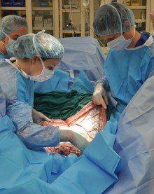
CALEC surgery represents a groundbreaking advancement in ocular trauma treatment, promising new hope for individuals suffering from blinding cornea injuries. This innovative procedure utilizes cultivated autologous limbal epithelial cells, a form of stem cell therapy, to restore the eye’s damaged corneal surface effectively. During a recent clinical trial at Mass Eye and Ear, this method demonstrated a remarkable 90 percent effectiveness rate in recovering corneal function over 18 months. The surgery involves harvesting healthy stem cells from one eye and transplanting them into the affected eye, allowing for the regeneration of the critical limbal epithelial cells that maintain a smooth corneal surface. As the first of its kind in the United States, CALEC surgery paves the way for transforming the lives of those with previously untreatable ocular conditions, offering a glimmer of hope for a clearer future.
Also referred to as cultivated limbal epithelial cell transplantation, CALEC surgery utilizes innovative bioengineering techniques to address severe injuries to the eye’s surface. This procedure is particularly vital for patients experiencing limbal stem cell deficiency due to trauma or disease. By leveraging stem cell therapy, clinicians can effectively restore the corneal surface, significantly improving visual acuity and alleviating persistent pain. As an exciting development in the realm of regenerative medicine, CALEC has opened avenues for novel treatments for ocular trauma, advancing strategies in how we approach the repair of corneal damage. The promising outcomes from ongoing studies could lead to wider adoption and improved protocols for treating patients suffering from debilitating visual impairments.
Introduction to CALEC Surgery: A Breakthrough in Eye Treatment
Cultivated Autologous Limbal Epithelial Cells, commonly referred to as CALEC, stand as a revolutionary advancement in ocular treatment for patients suffering from severe corneal damage. This surgery involves a meticulous process where stem cells from a healthy eye are cultivated and then transplanted into the affected eye, effectively rejuvenating the corneal surface. Introduced successfully in clinical trials at Mass Eye and Ear, CALEC surgery promises not only to restore vision but also to alleviate the chronic pain associated with blinding corneal injuries, marking a significant leap in ocular trauma treatment.
The efficacy of CALEC surgery is underscored by its impressive success rates observed in clinical trials, where over 90% of patients experienced substantial restoration of their corneal surface. This innovative procedure is particularly noteworthy due to its use of limbal epithelial cells, which are crucial for maintaining the integrity and transparency of the cornea. Prior to this development, patients suffering from limbal stem cell deficiency had limited options and faced bleak outcomes, highlighting the importance of continued research in stem cell therapy to enhance treatment accessibility.
How CALEC Surgery Works: The Process Explained
CALEC surgery is meticulously designed to ensure safety and efficacy in restoring the cornea’s surface. The process begins with the careful extraction of limbal epithelial cells from the patient’s healthy eye, which is accomplished through a minor biopsy procedure. These cells are then expanded into a cellular tissue graft under controlled conditions that meet rigorous quality standards. After a cultivation period of two to three weeks, the graft is surgically implanted into the damaged eye, facilitating the restoration of the cornea’s integrity.
This approach not only helps restore vision but also addresses the underlying issues of corneal damage caused by various factors, including chemical burns and infections. The ability to regenerate limbal epithelial cells through CALEC surgery provides a beacon of hope for patients previously left with few options. Moreover, the continuous improvements in the procedures and techniques surrounding CALEC highlight the ongoing dedication to improving outcomes for individuals suffering from ocular trauma.
Comparing Traditional Treatments and CALEC Surgery
Traditionally, patients with severe corneal injuries were often limited to treatments like corneal transplants, which may not be effective in cases of limbal stem cell deficiency. These standard procedures did not address the underlying damage and were often met with variable success. In contrast, CALEC surgery not only restores the cornea’s surface but also targets the specific problem at hand—the depletion of limbal epithelial cells. This method offers a more comprehensive solution than traditional options, particularly for patients facing chronic visual impairment.
The remarkable outcomes observed in the CALEC trials set it apart from conventional therapies. Instead of simply replacing the cornea, CALEC surgery empowers the body to heal itself by reinstating the vital cells that promote corneal health. The high success rates reported in the trials mean that more patients could potentially regain their vision and improve their quality of life significantly. This paradigm shift in treating corneal injuries shows that innovative approaches can yield better results than existing methodologies.
Clinical Trials and Results: The Evidence Behind CALEC
The clinical trial leading to the implementation of CALEC surgery at Mass Eye and Ear showcased its safety and feasibility extensively. Out of 14 participants, a staggering 93% showed signs of improvement within the first twelve months, proving that not only is this innovative procedure effective, but it is also safe for recipients. The structured follow-up and analysis further solidified CALEC’s standing as a groundbreaking treatment method, with no serious adverse effects noted during the trial.
The meticulous observation of participants over the course of 18 months revealed significant insights into the functionality and impact of CALEC surgery on visual acuity. Researchers noted varying improvements in vision among all patients, underscoring the procedure’s potential in reversing the debilitating impacts of corneal damage. Such results highlight the necessity for broader studies involving a larger patient demographic and longer-term assessments to ascertain the full scope of CALEC’s benefits.
Future Directions in Limbal Epithelial Cell Research
While CALEC surgery has demonstrated remarkable success, the vision for its future is even more expansive. Researchers are now focusing on establishing an allogeneic manufacturing process, which would allow stem cells to be harvested from cadaveric donor eyes. This innovation aims to enhance accessibility and provide treatment options for patients suffering from bilateral corneal injuries, who are currently excluded from receiving CALEC due to the reliance on a single healthy eye for stem cell extraction.
Fostering advancements in limbal epithelial cell therapy not only holds promise for expanding the potential patient pool but also pushes the boundaries of regenerative medicine in ophthalmology. The future of CALEC and similar therapies could pave the way for customized treatments developed from extensive research and collaboration among institutions, ultimately leading to a world where effective ocular trauma treatments are universally accessible.
Understanding Corneal Surface Restoration Through Stem Cell Therapy
The application of stem cell therapy for corneal surface restoration represents a significant advancement in addressing severe ocular conditions. At the heart of this treatment is the recognition that the deficiency of limbal epithelial cells leads to chronic corneal damage and visual impairment. By harnessing the regenerative capabilities of stem cells, CALEC surgery works to restore not just vision but the overall health of the eye, showcasing a new frontier in medical science.
This innovative approach bridges the gap between traditional surgical interventions and advanced cellular therapies. The ability to use a patient’s own cells to regenerate essential tissues within the eye harnesses the body’s innate healing processes, making it a potentially safer and more effective strategy than synthetic alternatives. As research continues to evolve, the integration of stem cell therapy into routine eye care practices may redefine the management of debilitating ocular disorders.
Addressing Ocular Trauma: The Role of CALEC Surgery
Ocular trauma remains one of the leading causes of blindness in various demographics. The potential impact of CALEC surgery in addressing these challenges is significant, offering renewed hope for recovery to thousands affected by traumatic eye injuries. By specifically targeting the restoration of limbal stem cells, CALEC represents a targeted approach to repairing the cornea and thus enhancing both functional outcomes and quality of life for affected patients.
The application of CALEC surgery in cases of ocular trauma is particularly compelling due to its dual benefits: not only does it aim to restore vision, but it also alleviates the persistent pain associated with corneal damage. This dual-action potential marks CALEC as a beacon of hope for victims of blinding cornea injuries. Moving forward, ongoing research and clinical trials will be critical in understanding the full capabilities of CALEC and its role within a broader context of ocular trauma treatment.
The Importance of Stem Cell Therapy in Regenerative Medicine
Stem cell therapy is emerging as a transformative approach across various fields of medicine, particularly in regenerative medicine. Specifically, for conditions affecting the eye, such as corneal injuries and diseases, the ability to utilize the patient’s own cells to regenerate damaged tissues signifies a new era of treatments. CALEC surgery exemplifies this innovation, revealing the profound potential of stem cells in restoring not only function but also quality of life for patients.
Offering hope to patients with conditions previously deemed untreatable, stem cell therapies like CALEC catalyze a shift from conventional surgical practices to a focus on healing and regeneration. This transition not only optimizes patient care but also encourages the exploration of new applications of stem cell technology in other aspects of medicine. As the field advances, continuous support for research and clinical trials will be vital to bring these innovative therapies to wider populations.
Funding and Support for Ocular Research Initiatives
The progression of CALEC surgery and other stem cell therapies owes much to the generous funding and support from organizations like the National Eye Institute. Funding ensures that researchers can pursue extensive studies that validate the safety and efficacy of these groundbreaking treatments. As CALEC surgery continues to evolve, the commitment from these institutions will be crucial in paving the path towards eventual implementation and accessibility of novel ocular therapies.
Furthermore, continued investment in ocular research initiatives amplifies the push towards not only understanding the complexities of eye health but also developing comprehensive treatment strategies that incorporate advances in cellular therapy. The collaborative efforts among institutions such as Mass Eye and Ear, Dana-Farber, and Boston Children’s highlight the synergistic benefits of both funding and research partnerships in fostering innovation in ophthalmology.
Frequently Asked Questions
What is CALEC surgery and how does it work?
CALEC surgery, or cultivated autologous limbal epithelial cell surgery, is a pioneering procedure that uses stem cells from a healthy eye to restore the corneal surface of a damaged eye. During the surgery, limbal epithelial cells are harvested from the patient’s unaffected eye and expanded into a graft over two to three weeks. This graft is then transplanted into the eye suffering from blinding cornea injuries, allowing for remarkable recovery and restoration of vision.
Who is a candidate for CALEC surgery?
Candidates for CALEC surgery typically include individuals who have sustained blinding cornea injuries, resulting in limbal stem cell deficiency due to conditions like chemical burns or ocular trauma. Patients should have only one affected eye, as a biopsy is required from the healthy eye to obtain the necessary limbal epithelial cells for the procedure.
What are the success rates of CALEC surgery for treating corneal injuries?
The clinical trial for CALEC surgery indicated a success rate of over 90% in restoring the corneal surface. Complete restoration was observed in 50% of participants at three months, which increased to 79% and 77% at 12 and 18 months, respectively. This demonstrates CALEC’s high potential in treating corneal surface abnormalities caused by severe injuries.
What is the role of stem cell therapy in CALEC surgery?
Stem cell therapy is at the core of CALEC surgery, as it involves cultivating limbal epithelial cells derived from a healthy donor eye to repair and regenerate the corneal surface of the damaged eye. This innovative approach offers new hope for patients with previously untreatable blinding cornea injuries by using the body’s own cells to promote healing.
Are there any risks associated with CALEC surgery?
While CALEC surgery has shown a high safety profile with no serious adverse events reported during the clinical trial, risks do exist. One participant experienced a bacterial infection associated with chronic contact lens use, though other minor adverse events were resolved quickly. As this procedure is still experimental, ongoing studies will help further assess its safety and effectiveness.
When will CALEC surgery be widely available to patients?
Currently, CALEC surgery is not offered at Mass Eye and Ear or any U.S. hospital, as it remains experimental. Additional studies and trials with larger patient groups are needed before the treatment can be submitted for federal approval for general use. Researchers are hopeful that further advancements will lead to broader accessibility for those suffering from corneal damage.
How does CALEC surgery differ from traditional corneal transplants?
Unlike traditional corneal transplants that rely on donors for corneal tissue, CALEC surgery utilizes the patient’s own limbal epithelial cells, minimizing the risk of rejection and complications associated with donor tissue. This personalized approach not only aims to restore vision but also addresses the underlying deficiency of limbal stem cells caused by ocular trauma.
What advancements are expected in the future of CALEC surgery?
Future advancements in CALEC surgery may include establishing an allogeneic manufacturing process that allows the use of limbal stem cells from cadaveric donors. This could potentially expand the treatment applications for patients suffering from bilateral corneal damage, thus broadening the impact of this regenerative therapy on ocular trauma treatment.
| Key Points |
|---|
| First CALEC surgery performed at Mass Eye and Ear by Ula Jurkunas. |
| Stem cell therapy developed to restore corneal surfaces in patients with damage. |
| Clinical trial involved 14 patients followed for 18 months with significant success rates. |
| Over 90% effectiveness observed in restoring cornea’s surface. |
| Procedure involves removal of stem cells from a healthy eye, expanding them, and transplanting them. |
| Current limitations require patients to have only one affected eye for stem cell sourcing. |
| Future research aims to establish allogeneic manufacturing to help patients with bilateral eye damage. |
| Procedure showed safety with no serious adverse events reported. |
| Additional studies needed before broader application and FDA approval. |
Summary
CALEC surgery heralds a groundbreaking advancement in the treatment of corneal injuries once deemed irreparable. Through innovative stem cell therapy, this pioneering procedure has shown remarkable efficacy, achieving over 90% success rates in restoring damaged corneal surfaces. With an encouraging safety profile and potential for broader application, CALEC surgery offers new hope to patients suffering from visual impairments due to corneal injuries.

